How to Describe an Abscess on Physical Exam
Later as the abscess points the overlying skin becomes thin and feels fluctuant. Was this post helpful.

Skin Abscess Causes Diagnosis And Treatment
Inspection palpation and lymph node exam.
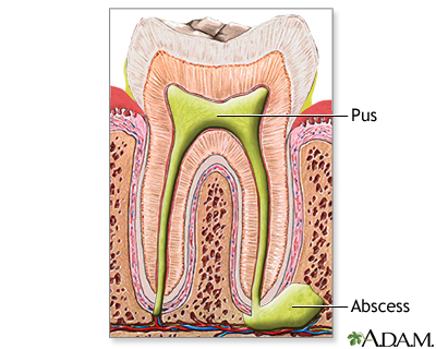
. Signs and symptoms include the presence of a swollen tender and erythematous nodular lesion in the skin associated with fever and chills. Cutaneous abscesses are painful tender indurated and usually erythematous. Characteristic findings on physical examination On physical examination they are characterized as fluctuant subcutaneous collections with overlying erythema and edema Figure 1.
A skin abscess is round and feels firm and squishy due to the thick membrane around it and the liquid pus inside. The size of the tonsils has no bearing on the presence or absence of a peritonsillar abscess. Use the flat of your outstretched right hand with the thumb tucked under the palm placed at right angles to the costal margin.
Wearing gloves may reduce your ability to fully appreciate all the features of a. The abscess may then spontaneously drain. Press with one finger and feel whether the lump bounces against your other finger.
Causes include folliculitis furuncle skin injury and bacterial infections. These abscesses can cause significant discomfort for patients. Possible drainage if there is an infection.
Initially the swelling is firm. A health care provider may mark the edges of the redness with a pen to see if the redness goes past the marked border over the next several days. To test for fluctuation put your fingers on either side of the lump opposite each other.
Cluster1homenancyclark1 TrainingEMRSOAP Notedoc O. Listed are the components of the all normal physical exam General. It is most commonly a manifestation of a staphylococcal infection.
An abscess is a localized cavity filled with puss. This will involve me using a. Up to 6 cash back A cyst is a bag-like structure lined by a membrane and filled with fluid or a semi-fluid material.
An untreated peritonsillar abscess can lead to oropharyngeal obstruction and trismus due to odynophagia. Staples are intact with some erythema that is a reaction to the staplessutures. Dental caries are caused by the following.
Muffled voice also called hot potato voice Contralateral deflection of the uvula see image below. The American Academy of Pediatrics AAP along with the American Academy of Pediatric Dentistry issued a clinical. By the way local or localized means that the swelling is not systemic in other words it doesnt affect the whole body.
Underneath the skin it may create a swollen bump. They can develop on top of the skin under the skin in a tooth or even deep inside the body. Generally if a wound looks good it means there is no evidence of infection or dehiscence gaping open and that it is healing.
A hematoma is a localized cavity occupied by a blood clot. Press the radial border of your index finger into the abdomen during expiration and retain in this position. Intermittent hot compresses are used to facilitate drainage.
An abscess that develops in the skin and subcutaneous tissues. Swollen glands lymph nodes near the affected area. Abscesses are often easy to feel by touching.
Abscesses are incised and drained. The physical exam of the breast can be divided into three components. They are usually acutely ill-looking.
The term early childhood caries is replacing these terms because the description also includes dental caries in breastfed babies. Often the complication occurs when the tonsils are barely enlarged. If the lump is thought to contain fluid this can sometimes be confirmed by eliciting a fluid thrill.
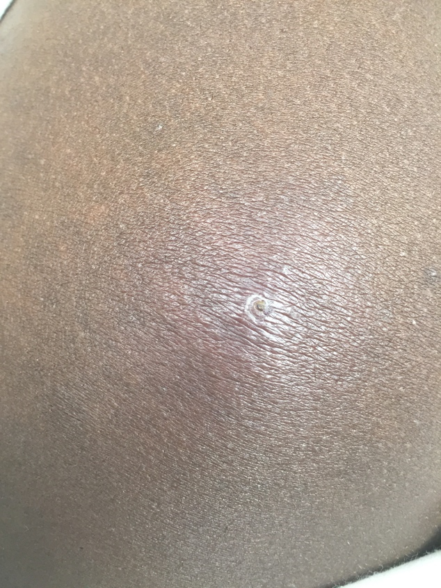
They vary in size typically 1 to 3 cm in length but are sometimes much larger. On top of the skin an abscess might look like an unhealed wound or a pimple. Oct 29 2005.
Infant-bottle tooth decay or nursing caries. The vast majority of them are caused by. Abscesses are commonly located in the axillae groin and rectal area but can be located in any area.
In performing the breast exam is important to keep in mind the following general points. Antibiotics when used should be effective against MRSA MRSA and purulent or complicated cellulitis Cellulitis is acute bacterial infection of the skin and subcutaneous tissue most often caused by streptococci or staphylococci. They are located at the anal verge and if left untreated can extend into the ischioanal space or intersphincteric space since these areas are continuous with the perianal space.
Sometimes there is a pinpoint opening in the center a punctum. Good turgor no rash unusual bruising or prominent lesions Hair. Redness warmth and swelling of the skin may be present.
Explain examination and obtain consent. - It is better not to wear gloves while palpating the breasts. Streptococci abscess Fluid Fluid filled abscess Friable Clostridial hepatopathy Gritty Urinary calculi Leathery Chronic mange Resilient The normal nose Rubbery PRRSv in lungs Spongy Udder oedema Viscous Shoulder abscess.
This indicates a fluid- or fat-filled lump. - for stapled or sutured wounds. Persistent abdominal pain focal tenderness spiking fever persistent tachycardia prolonged ileus leukocytosis or intermittent polymicrobial bacteremia suggest an intra-abdominal abscess in.
Physical examination findings suggestive of peritonsillar abscess include the following. Wound looks good with no drainage. Abscess Overview A skin abscess is a tender mass generally surrounded by a colored area from pink to deep red.
Symptoms and signs are pain warmth rapidly. There are several ways of describing them. Well appearing well nourished in no distressOriented x 3 normal mood and affect.
Abscesses usually are red swollen and warm to the touch and might leak fluid. Normal texture and distribution. Perianal abscesses are the most common type of anorectal abscesses.
It is usually painful and the overlying skin is often red. Appearance of the Patient. A large liver extends down towards the right iliac fossa and the edge is often firm and easily palpable.
Abscess Information Mount Sinai New York
Abscess Incision And Drainage

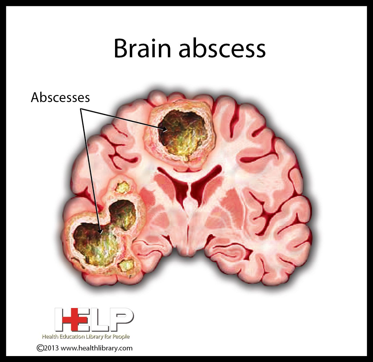
Brain Abscess Nursing School Studying Nursing School Notes Nursing School Survival

Skin Abscess Causes Diagnosis And Treatment

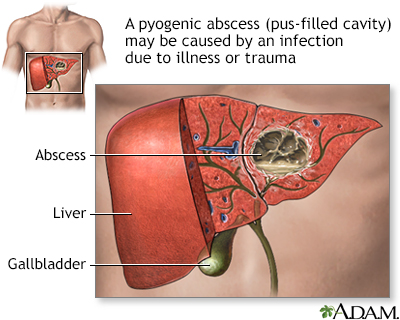
Pin On Digestive System

Skin Abscess Causes Diagnosis And Treatment
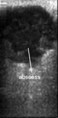
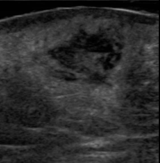
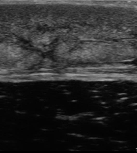
Abscess Evaluation With Bedside Ultrasonography Practice Essentials Technique Pearls

Skin Lesions Nursing Study Tips Skin Assessment Nursing Information
/lung-abscess-overview-4768089_final-8c6a0ccd797345289aa2dc0a5856d8e1.jpg)
Lung Abscess Symptoms Causes Diagnosis And Treatment

Image Result For Retropharyngeal Abscess Anatomy
2

Pelvic Inflammatory Disease And Tubo Ovarian Abscess Pelvic Inflammatory Disease Endometrial Biopsy Ovarian
Abscess Information Mount Sinai New York

Department Of Surgery Perianal And Perirectal Abscess Fistula
Abscess Incision And Drainage
Abscess Incision And Drainage

View More Teaching Images And Get Your Free Trial At Roshreview Com Cauda Equina Physical Therapy School Medical Mnemonics
Brain Abscess Nclex Nurse Practioner Nursing Students
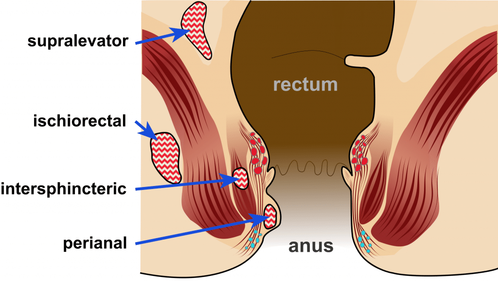
Anorectal Abscess Risk Factors Management Teachmesurgery


Comments
Post a Comment